
-composed of 206 bones and the articulations between the bones
Functions
1. Support
2. Protection---internal organs
3. Movement facilitation---
-bones act as the levers
-articulations act as the fulcrums
-muscles provide the force
4. Mineral Storage---calcium and
phosphorus
5. Storage of Blood Cell Producing
Cells---red bone marrow
6. Storage of Energy---lipids stored in
yellow bone marrow are important source of chemical energy
Cells and Histology of
bone
-skeletal system consist of cartilage, bone and dense connective
tissue
4 types of cells in bone (osseous)
tissue
1. Osteoprogenitor---
-found throughout the bone
-have mitotic potential
-may differentiate into osteoblast
2. Osteoblast---
-no mitotic potential
-found on the surface of the bone
-secrete mineral salts and organic components for bone formation
3. Osteocytes---
-no mitotic potential
-found within the bone
-maintain daily cellular activities of bone tissue
4. Osteoclast---
-found on the surface of bone
-function in the reabsorption of bone
1. Diaphysis---
-shaft or main part of the long bone
2. Metaphysis---
-area between diaphysis and epiphysis in mature bone
3. Epiphysis---
-ends of the bones
4. Articular
Cartilage---hyaline cartilage
5. Periosteum---
-contains nerves, blood vessels, osteo cells
-serves as point of attachment for ligaments and tendons
6. Medullary or Marrow
Cavity---contains yellow marrow
7. Endosteum---lines the
medullary cavity
Spongy
Bone---
-consists of an irregular lattice work of bone called
trabeculae
-spaces are filled with red
marrow---responsible for producing red blood cells
Compact Bone---
-tightly packed tissue
-parts of compact bone
Concentric Lamellae
-Concentric circles of tightly packed tissue
Volkmann’s Canals
-penetrate the compact bone
-allow blood vessels and nerves to go to the medullary cavity and
other Haversian Canals
-run perpendicular to the long axis of the bone
Haversian Canals---
-run longitudinally through the compact bone
-concentric lamellae surround the canals
Lacunae---
-open spaces between the concentric lamellae that contain
osteocytes
-look like “little lakes”
Canaliculi---
-tiny canals that radiate away from the lucunae
Osteon (Haversian
System)
-central canal, surrounding lamellae, lacunae, osteocytes and
canaliculi

- bone development
1. Hyaline
Cartilage
“bone” created by chondroblast
-actual ossification of this “bone” starts 6-7 weeks after
conception
2. Interstitial and Oppositional
Growth
-chondrocytes burst and this triggers calcification
-dying cartilage is invaded by capillaries and bone cells
-bone starts to calcify
3. Primary Ossification Center
Develops
-blood vessels in mid-region produce ossification center
-osteoblast for spongy bone in the area of calcified cartilage, this
causes the center to enlarge
-this spongy bone is destroyed by osteoclast, leaving the medullary
cavity
4. Secondary Ossification
Center
-when blood vessels enter the epiphysis
-spongy bone created here will stay
- epiphysial plate---
-layer of hyaline cartilage between the 2 areas of growth
-stays until you reach maturity
5. Remnants of Hyaline
Cartilage on the epiphysis is the articulating cartilage

1. Synarthroses
(juncture fibrosae)
-bones connected by fibrous tissue or cartilage
-processes are interlocked (NO movement) (ex. between skull
bones)
2. Amphiarthroses
(juncture cartilagenae)
-slightly movable (ex. pubic symphysis)
3. Diarthroses
(juncture synoviales)
-freely movable
-most joints of the body
-end of the bones covered in hyaline cartilage and surrounded by a
fibrous capsule
-stabalized by ligaments and tendons that pass over the fibrous
capsule
-also called synovial joints because of the synovial fluid found in
the capsule that reduces friction of the articulation
-classified by the kind of motion that they permit
- A) Gliding
joints
-ends of bones glide over each other
-articular surfaces are almost flat (ex. bones of wrist and
ankles)
B) Hinge
joints
-allow angular motion in ONE direction
-convex surface of one bone fits into the concave surface of the
other
-allows for flexion, extension, and sometimes hyperextension (ex.
elbow, knee and ankle)
C) Condyloid joints
-angular movement in TWO directions
-oval shape condyle of one bone fits into a cavity or fossa of the
other
-capable of circumduction, flexion, extension, abduction and
adduction (ex. articulation between the carpals and the radius and
ulna)
D) Saddle
joint
-allows for the same movements as condyloid joints (ex.
thumb and trapezium)
E)
Pivot joints
-pointed process of one bone turns within a ring formed
-partly by another bone and partly by a ligament
-allows for rotation (ex. atlas and axis)
F) Ball and socket
joints
-have angular movement in all directions
-rounded head of one bone lies in a cup like cavity of another
(ex. hip and shoulder--the most freely movable joint in the
body)
Types of articular movements
1. Gliding--surface of one bone moves
over the surface of another bone
-simplest type of joint movement (ex. between the tarsals)
2. Angular movements
-occurs only when the angles of the bones are affected
- a) Flexion--decrease in the angle
between the bones
b) Extension--angle is increased
c) Abduction--drawn away from the
midline of the body
d) Adduction--bring toward the
midline of the body
- 3. Rotation--bone moves around a
central axis
4. Circumduction--the distal end of a
bone moves around a circle while the proximal end remains
stationary
-the bone outlines a “cone” in the air
5. Inversion--sole of the foot turns
toward the midline
Eversion--sole of the foot turns away
from the midline
6. Special movements
a) Protraction--move forward on a
plane parallel to the ground
Retraction--move backward
b) Supination--turns the palm
anterior
Pronation--turns the palm
posterior
c) Elevation
Depression

Process--bony
prominence “bump”
1. Condyle-- rounded or knuckle like
process
2. Tubercle-- small process
3. Tuberosity-- large process (tibial
tuberosity)
4. Trochanter-- huge process (located on
the proximal end of the femur)
Crest--narrow ridge
of bone
Spine--sharp slender
process (Holes and / or depressions)
Fissure--narrow slit
through which blood vessels or nerves pass
Foramen--opening
through which blood and nerves pass
Meatus--tubelike
passageway running within a bone
Sulcus or
groove--furrow that accommodates a soft structure such as
blood vessels, nerves or tendons
Fossa--depression in or
on the surface of a bone

15 types of fractures
1. Partial
2. Complete
3. Closed
(simple) -doesn’t break the skin
4. Open
(compound) -breaks the skin
5. Comminuted
-bone splinters at the site of the impact
6. Greenstick
-partial break where one side breaks and the other side bends
7. Spiral -break
by being twisted
8. Transverse
-fracture at right angles to the long axis of the bone
9. Impacted -one
fragment is firmly driven into the another
10. Displaced
-anatomical alignment is not preserved
11.
Non-displaced
-anatomical alignment is preserved
12.
Stress-partial fracture
resulting in bones inability to withstand forces
-about 25% involve distal end of the fibula
13. Pathologic
-caused by weakening of the bone due to a disease
14. Pott’s
-fracture of the distal end of the fibula with serious injury to the
distal tibial articulation
-severe eversion sprain may lead to this
15. Colle’s
-fracture at the distal end of the radius in which the distal ends is
displaced posteriorly
-occurs frequently when you try to stop yourself from falling

-fracture breaks blood vessels found in the Haversian System
-blood clot forms at the site of the break within 6 to 8 hours
(fracture hemotoma)
Fracture
hemotoma---serves as the focus for cellular invasion
Callus--
-new bone tissue developed around the fracture
-site of osteoblast activity
Remodeling--
-dead bone is absorbed by osteoclast
-compact bone replaces spongy bone in the fractured area

Osteoporosis--
-age related disease characterized by decreased bone mass and an
increased chance of fractures
-decreased levels of
estrogen (sex hormones
that stimulates osteoblast)
-white women are affected more than men and people of color
-other factors linked to
osteoporosis
- a) body build-short people
have less bone mass
b) weight-thin people have
less adipose which stores estrogen
c) smokers-low estrogen
levels
d) calcium deficiency and
malabsorption
e) vitamin D deficiency
f) certain
drugs-alcohol, cortisone, etc.
g) premature menopause
Steps to Prevent
Osteoporosis
- Exercise--muscle action
stimulates blood flow to bone tissue
Estrogen Pills-for post
menopause women
Calcium supplements /
vitamins
Prescription anabolic
steroids-increase hormone levels
Sodium
fluoride-stimulates osteoblast
Osteogenic Sarcoma--
-malignant bone tumor that effects osteoblast
-people between ages of 10-25
-left untreated it will metastasize and kill you
-treatment includes chemotherapy following amputation of affected
area
Rickets--
-vitamin D deficiency in children
-body can’t transport calcium and phosphorus from the G.T. tract
into the blood
-osteoblast in diaphysis don’t calcify which causes the bones to
stay soft
-weight of body causes legs to bow
cure and prevention
-dietary--add vitamin D, calcium and phosphorus in great amounts
-exposing the skin to ultraviolet rays of light

ARTHRITIS--
-refers to many diseases characterized by an inflammation of one or
more joints
- Rheumatoid
Arthritis--
-auto immune disease in which the body attacks its own tissue, in
this it attacks cartilage and joint linings
-primary symptom is inflammation of the synovial membrane
-treatments aimed to reduce pain and inflammation while preserving
strength and mobility (rest, aspirin, steroids, exercise,
etc.)
-likely to be bilateral in areas affected
-usually affects small joints 1st
Osteoarthritis--
-more common and less damaging
-deterioration of articular cartilage and formation of bone in the
joint (bone spurs)
-non inflammatory, progressive disorder
-bone spurs decrease articular cavity and restrict movement
-effects large weight-bearing joints
-results from a combination of aging, irritation of the joint, and
normal wear and tear
-treatments are similar to Rheumatoid Arthritis
Gouty Arthritis--
-gout is a disease when your body produces excess amounts and/or
is not able to excrete normal amounts or
uric acid-- waste produced
when nucleic acid is metabolized
-excess uric acid reacts with sodium to form a salt called sodium
urate
-sodium urate crystals are deposited into soft tissues (articular
cartilage, kidneys, ears)
-crystals irritate and wear down the cartilage eventually
destroying all joint tissue and wearing down bones
-if not treated the bones might fuse and become immovable
-effects primarily middle to older males
-treatment can be successful (other two types cannot be fully
treated) by controlling the uric acid production

Comprised of 2 parts
pectoral girdle and upper extremity
pelvic girdle and lower extremity
Pectoral
Girdle
-function--attach bones of the upper extremities to the axial
skeleton
-composed of the clavicle on the anterior and the scapula on the
posterior
Clavicle--
-long slender bone with a double curvature
- sternal end--
rounded end that articulates with the sternum
acromial end--
broad and flat end the articulates with the acromian of the
scapula
Scapula--
-triangular flat bone found on the posterior part of the thorax
between the levels of the 2nd and 7th rib
-medial boarder is about 5 centimeters from the vertebral column
- spine-- runs diagonally across
the body
acromian process-- is part of the
spine that expands past the body
glenoid cavity-- fossa inferior to
the acromian process. Articulation with the head of the humerus to
make a ball and socket joint.
supraglenoid tubercle and infraglenoid
tubercle-- help form the socket
coracoid process-- located at the
lateral end of the superior ridge and it projects on the anterior
side
scapular notch-- predominant
indentation of the superior ridge next to the coracoid process
Upper
Extremities
-consist of 60 bones
-each side contains 30 bones (1 humerus, 1 radius, 1 ulna, 8 carpals,
5 metacarpals and 14 phalanges)
Humerus--
-articulates proximally with the scapula and distally with both the
radius and the ulna
-largest bone in the upper extremity
- head--rounded proximal end,
articulates with the glenoid cavity
anatomical neck--
-slight groove found just below the head
-epiphysial plate found in this area
greater tubercle--large. lateral
process found just below the anatomical neck
lesser tubercle--found on the
anterior side
intertubercular groove
surgical neck--
-constricted portion, inferior to the tubercles
-fractures likely to happen here
deltoid tuberosity--
-slight bump on the anterior surface of the humerus
-insertion point of the deltoid muscle
capitulum--rounded knob that
articulates with the head of the radius
trochlea--pulley like surface that
articulates with the ulna
medial and lateral epicondyles--
-found on either side on distal end of the humerus
-serve as attachment of most of the forearm muscles
coronoid fossa--
-receives part of the ulna when elbow is flexed
-on anterior, distal surface
olecranon fossa--posterior side and
receives olecranon process of the ulna when elbow is extended
ulnar nerve--
-lies over the posterior surface of the medial epicondyle
-can be rolled between the finger and the medial epicondyle
Ulna--
-medial bone of the forearm
- olecranon process--posterior
projection on the proximal end of the ulna
coronoid process--anterior
projection
trochlear (semilunar) notch--
-curved area between olecranon and coronoid processes
-articulates with trochlea of humerus
-allows for flexion and extension of the elbow
styloid process--posterior side of
the head of the ulna
head--distal end of the ulna
Radius--lateral bone
found on the thumb side of the forearm
- head--
-disc like structure that articulates with the capitulum of the
humerus and the radial notch of the ulna
-attachment point of biceps brahii muscle
Carpals,
Metacarpals and
Phalanges
Carpals--
-eight small bones that are connected by ligaments to form the wrist
(carpus)
-arranged in 2 transverse rows, 4 bones per row
-named for their shapes (proximal row from lateral to medial)
- scaphoid--
-resembles a boat
-70% of wrist fractures
lunate--looks like a crescent
moon
triquetrum--has 3 articular surfaces,
looks like a triangle
pisiform--pea shaped, very small
(distal row from lateral to medial)
trapezium--4 sided bone that
articulates with the thumb, saddle shaped bone
trapezoid--4 sided bone articulates
with the proximal end of 2nd metacarpal
capitate--largest of the carpals that
articulates with lunate and the 3rd metacarpal
hamate--large hook shaped projection
on its anterior surface
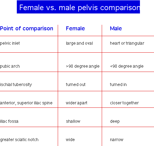
Pelvic Girdle
-2 coxal (hip) bones and sacrum for the pelvis
-provide a strong support for the lower extremities
Os
Coxa--
-one hip bone
-each made of 3 bones fuse together
1. Ilium--
-largest of the 3 subdivisions
-superior to the other 2 portions
- iliac crest--superior border that
serves as an insertion point for muscles of the abdominal wall
iliac fossa--internal surface seen
from the medial side that gives the pelvis a bowl shape
appearance
greater sciatic notch--
- -inferior to iliac crest and posterior to the acetabulum
-allows for major nerves and blood vessels to travel from your
sacrum to your legs
acetabulum--lateral fossa of the os
coxa where 3 pelvic bones merge here
- 2. Pubic
Bone--
-anterior, inferior part of the os coxa
-joins with the other pubic bone at the pubic symphysis
**hormone, relaxin, is released during childbirth that allows
greater flexibility of the fibrocartilage.
-
- orbturator foramen--between
ischium, pubic bone and acetabulum
- 3.
Ischium--inferior,
posterior portion of the coxal bone
-
- ishial tuberosity--tuberosity
that we sit on
Lower Extremity
each contains 30 bones
Femur--
-largest and heaviest bone in your body
-body of femur angles toward the midline makes knees closer than
hips
-degree is greater in females
- head--rounded end that
articulates with the acetabulum
neck--
- -distal to the head
-elderly people break this area often
greater and lesser
trochanters---large processes below the neck and serve as
points of attachment for some of the thigh and buttock muscles
shaft or body--diaphysis of femur
linea aspera---found on posterior
side of the shaft serves as an insertion point for adductor
muscles of the leg
medial and lateral condyles--distal
end of femur which articulates with the proximal end of tibia
intercondylar fossa--important area
for the ligaments in the knee
patellar surface--distal anterior
surface between the condyles that forms a gliding joint with the
patella
Tibio-femoral
Articulation
-largest joint in the body
-comprised of 3 joints (1 for each condyle and 1 between patella and
femur)
- 1. Synovial capsule
2. Patellar ligament--
-central portion of insertion for the quadriceps muscles
-strengthens anterior surface
3. Popliteal ligaments--crisscross
ligaments on the posterior surface
4. Medial (tibial)
collateral---provides lateral support and is easily
injured
5. Lateral (fibular) collateral
6. Intra articular ligaments
a) posterior cruciate--keeps tibia from sliding backward (back
part of “X”)
b) anterior cruciate--keeps tibia from sliding forward (anterior
part of “X”)
7. Meniscus--fibrocartilage that may
tear and the loose parts may impede movement
8. Bursae---sac of synovial fluid
found at the friction points of your body
TERRIBLE TRIAD
-tear in the medial collateral, anterior cruciate and medial
meniscus
causes--sports or
accidents
treatment--surgery with
intense rehabilitation
Patella
-”knee cap”
-inferior end is called the
Apex
-posterior surface has 2 articulating surfaces
Tibia
-”shin bone”
-large, medial, weight bearing bone of the lower extremity
-lateral and medial condyles articulate with the condyles of the
femur
-inferior surface of the lateral condyle articulates with the
fibula
- Tibial Tuberosity
-anterior surface of the tibia
-surface serves as a point of attachment for the patellar
ligaments and tendon
Intercondylar Eminence
-upward projection found between the condyles of your tibia
-cruciate ligaments attach here
Medial Malleolus
-medial projection on the distal end of the tibia
-provides medial support to the hinge joint of your ankle
Fibula--lateral, non-weight
bearing bone in your lower extremity
- Head--proximal end, more rounded
of the 2
Lateral condyle
-distal end that articulates with the tibia and talus
-lateral support to the “ankle”
-longer “pointier” end
(Pott’s Fracture)
Tarsals
-comparative to carpals
-only 7 tarsals
- Talus
-uppermost tarsal
-only bone that articulates with the tibia and fibula
-initially bares all of the body weight, then transfers one half
the the calcaneous and one half to the other tarsals
Calcaneous
-largest and strongest tarsal
-supports body weight
-serves as the lever attachment for the gastrocnemius
Navicular--found just anterior to the
talus
Cuboid
-found on the lateral side of the foot
-articulates with the calcaneous and the IV and V metatarsal
Cuneiforms
-”wedge shaped”
-numbered from the medial to the lateral side
-3 separate bones that each articulates with the corresponding
metatarsal
- Metatarsals--numbered
from the medial to the lateral side
Phalanges--similar to the
upper extremity
Arches of the foot
-enable the foot to support weight
-provide leverage while walking
-have some “spring” to them
-transverse and
longitudinal arch
-