
-skin and its derivatives such as hair, nails, glands and
specialized receptors
-occupies surface area of about 2 square meters
-Dermatology---medical specialty that diagnoses and treats skin
disorders

Physiology
1. Regulate Body Temperature---sweat and changes of blood flow
toward the surface of the skin
2. Protection---from abrasions, bacteria, dehydration and
ultra-violet radiation
3. Reception of Stimuli---nerve endings specifically designed for
pressure, temperature, touch and pain
4. Excretion---sweat helps to reduce levels of water, salts and
other organic compounds
5. Synthesis of Vitamin D---helps in the manufacturing of Vitamin
D which helps the body to absorb calcium and phosphorus from food
6. Immunity---certain cells help in your ability to produce
antibodies

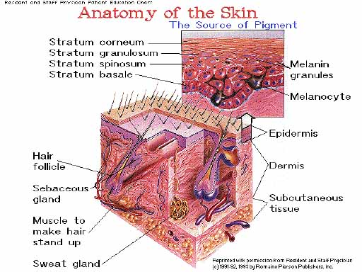
2 general structures plus epidermal
derivitives

-outer, thinner layer composed of keratinized stratifies squamous
epithelium
-4 special cells found here
- A) keratinocyte---produce keratin
- B) melanocyte---
- -found at the base of the epidermis
- -produce
melanin---responsible for skin
color and the absorption of ultra-violet radiation
- C) Langerhan's Cells---interact with helper T-cells of the
immune system
- D) Granstein Cells---
- -resistant to ultra-violet radiation
- -interact with other T-cells from the immune system
LAYERS OF THE EPIDERMIS
- 1. Stratum Basale---
- -single layer of cells capable of continued cell division
- -also called the stratum germinativum
- -cells may migrate to the dermis to become glands and hair
follicles
- -areas with no hair contain nerve endings that are sensitive
to touch (tactile disc)
-
2. Stratum
Spinosum---
- -8 to 10 rows of close fitting cells
- -surface of the cells contain spine like projections that help
to join cells together
-
3. Stratum
Granulosum---
- -3-to 5 rows of flattened cells that contain dark stained
granules of keratohyalin which is involved in the first steps of
keratin formation
- -cells start to die in this layer
-
*4.* Stratum
Granulosum---
- -found ONLY in thick shin of palms, and the soles of your
feet
- -3 to 5 rows of clear, flat, dead cells
-
5. Stratum
Corneum---
- -25 to 30 rows of keratinized cells
- -continuously replaced and shed

-consist of collagenous fibers and elastic fibers of connective
tissue
-blood vessels, nerves, glands, and hair follicles are found
here
-2 layers thick
1. Papillary Layer---
-upper 1/5 of the dermis
- -dermal papillae---
- -finger like projections that increase surface area
- -projections that extend into the epidermis and may contain
blood vessels or
- meissner's corpuscles---
endings that are sensitive to touch
- -cause ridges in the overlying epidermis (fingerprints)
2. Reticular Layer---
- -formed by closely packed irregularly arranged connective
tissue
- -spaces between are filled by glands, hair, and nerves
- -provides the skin with its strength and elasticity
- -attached to underlying organs by the subcutaneous layer
- Pacinian
Corpuscles---
- -located in the subcutaneous layer
- -nerve endings that are sensitive to pressure

- -develop from embryonic epidermis
- -pili

- -protects--guards scalp from the sun, eyes from foreign
particles
- -hair in the ear and nose protect these structures from
foreign particles and insects that might be inhaled or crawl into
the ear
Anatomy of the Hair
A) Shaft---
- -superficial portion that most of which projects above the
skin
- -made up of 3 parts
-
- 1. Medulla---inner portion
that contains air spaces
- 2. Cortex---
- -middle portion that makes the majority of the hair shaft
- -contain pigment granules of dark hair and mostly air in light
hair
- 3. Cuticle---
- -outer most layer
- -cells heavily keratinized
- -arranged like upside-down shingles
B) Root---
- -portion that penetrates the dermis and even the
subcutaneous
- layer
- -contains the 3 portion like the shaft
C) Hair follicle---
- -surround the root
- -continuation of the stratum basale and stratum spinosum
layers of the epidermis
- -base of each follicle enlarges and looks like an onion shaped
bulb
D) Papillae of the hair---
- -indentation of the bulb that is filled with loose connective
tissue
- and many blood vessels
Associates of the Hair Complex
1. Sabaceous (oil)
glands---
- -found in association with hair follicles except on the lips
and eyelids
- -secrete sebum---
- -mixture of fats, cholesterol, proteins, and inorganic
salts
- Function
- -prevents hair from becoming brittle
- -forms a film that prevents excess evaporation of water from
the skin
- -keeps the skin soft and flexible
- -inhibits the growth of certain bacteria
Clinical Application:
Blackheads
- -sebaceous glands enlarge due to accumulated sebum
- -color of blackhead is due to melanin and oxidized oil, not
dirt
2. Suderiferous Glands---
- -divided on basis of structure and location
- -3 types
- A) Apocrine---
- -located in axilla and pubic region
- -excretory duct open to hair follicles
- -start to function at the onset of puberty
- -omit an odor
- B) Eccrine---
- -more common
- -ducts open to the surface of the skin
*both of these types of glands secrete
- perspiration--- mixture of
water, salts (NaCl), urea, uric acid, amino acids, ammonia, sugar,
lactic
- acid and ascorbic acid
-
Function:
- -reduces body temperature by evaporation
- -elimination of waste
-
C) Mammary
glands---
- -modified suderiferous glands
- -reproductive unit
-
- D. Ceruminous
glands---
- -found in external auditory meatus
- -produce ear wax that protect your ears from foreign
particles

- -average growth is about 1 mm per week
- -protect the end of the digits and aid in the manipulation of
small objects
4 parts of a nail
1. Free Edge---sticks out past
distal end of the digit
- 2. Body---
- -majority of the visible nail
- -lunula---semilunar whit par of the body
3. Nail Root---hidden part of
the nail that lies above the nail matrix
- 4. Nail Matrix---function
to bring about the growth of nails when superficial cells
- become nail cells, they push the whole nail across the
nail
- bed

Common skin wounds
Abrasions---portion of the
skin has been scraped away
Lacerations---irregular tear
of the skin
Puncture---hole "popped"
through the skin
Incisions---clean cut through
the skin
- Contusion---
- -bruise
- -tissue below skin damaged, but skin is not broken
Superficial Wound Healing
1. Basale cells in the area of the wound break contact with the
basement membrane, that connects the epidermis to the dermis
2. Basale cells enlarge and migrate
across the wound
3. Contact inhibition stops the
migrating cells and turns cells in a new direction Continues until
cells are surrounded by similar cells.
**malignant (cancer) cells don't follow the same rules they
continue to spread and invade other areas
- 4. When the "Floor" of the wound is covered
- -cells divide to form new strata (layers)
- -this thickens the epidermis and fills in the wound from the
bottom upward
5. If a scab was formed, it will fall off when the new epidermis
is thick enough to protect itself.
- Deep Wound
Healing
- -when the injury extends past the epidermis
- -commonly due to accidental lacerations or surgical
incisions
- -scar formation will occur
- -repair more complex (4 phases)
-
1. Inflammatory
phase---
- -inflammation is the vascular and cellular response to rid
wound of bad stuff
- -blood clot forms to keep the edges of the wound close
together
- -epidermal cells start to migrate
- -vasodilation floods the area with phagocytic cells and
fibroblast
-
2. Migratory
phase---
- -clot becomes scab and epithelial cells continue migrating and
bridge the wound
- -fibroblast synthesize scar tissue
- -damaged blood vessels begin to regrow
-
3. Proliferative
phase---
- -epithelial cells grow under the scab
- -collagenous scar tissue is deposited
- -continued blood vessel growth
-
4. Maturation
phase---
- -scab falls off
- -collagenous fibers become more organized
- -blood vessels restored to normal
- -scar tissue---
- -dense collagenous fibers
- -fewer blood vessels
- -may not contain hair, glands, or sensory receptors

- 1. Melanin---
- -pigment found primarily in the basale and spinosum layers of
the epidermis
- -varies skin color from yellow to black
- -number of melanocytes about the same for all races
- -skin color due to the amount of pigment the melanocytes
produce
-
Disorders
- Albinism---inability to produce melanin
- Vitiligo---loss of melanocytes from an area of skin
- Freckles---patches of melanin
- Tanning---
- -also associated with the melanin
- -ultra-violet radiation increases melanocyte activity
-
2. Carotene---
- -found in the stratum corneum
- -people of Asian origin have carotene in fatty areas of the
dermis
- -gives a yellowish hoe to the skin
-
-
3. Capillaries---cause the
skin to have a pink appearance
- Disorders
- Malignant Melanoma
- -cancer of the melanocytes
- -overexposure to ultra-violet light of the sun may causes
- -most tumors involve basale cells, so they can be removed
surgically
-
BEST TREATMENT IS PREVENTION
- -examine your skin for moles that develop irregular
borders,
- uneven surfaces, or a mixture of colors or change in size or
start to bleed
- -any of these may be a sign of a developing melanoma
-
Wrinkles
- Collagen fibers---stiffen, break apart and form a
shapeless tangle
- Elastic fibers-- some elasticity, thicken and fray
- Subcutaneous fat---decreases
- Sebaceous glands---atrophy leads to dry, cracked
skin
-
other problems:
- Melanocytes---
- -decrease of functioning melanocytes lead to gray hair and
atypical skin pigmentation
- -increase of size of some melanocytes can cause liver
spots
- -older skin more susceptible to pathological conditions like
cancer and senile pruritis (itching)
-
Cystic
Acne---
- -scarring as a result of severe acne
- -surrounding epidermal cells might be replaced by connective
tissue
- -squeezing, pinching or scratching the lesion will increase
the chance of developing cystic acne
-
Psoriasis---
- -symptoms are distinct, reddish, small round skin elevations
covered with scales
- -caused by abnormally high rate of mitosis in the epidermal
cells
- -trauma, infection, stress, seasonal or hormonal changes can
initiate
- -treatments include steroid ointments and natural sunlight
(ultra-violet light)
-
Sunburn---
- -dead layers of cells peel off and leave unprotected layers of
cells
- -ultra-violet rays of sunlight damage the cell's DNA and
RNA