1. SENSORY
-senses changes in and outside the body
2. INTEGRATIVE
-interprets these changes
3. MOTOR
-responds to the changes by initiating action in the form of muscular
contractions or glandular secretions
* most rapid means of maintaining homeostasis in your body
* study of the nervous system is called neurology
![]()
2 Main divisions
1. CENTRAL NERVOUS SYSTEM (CNS)
-brain and spinal cord
-all sensations have to be relayed here to be acted on
-muscle and gland stimulation
-control center for the entire system
2. PERIPHERAL NERVOUS SYSTEM
(PNS)
-connection between the CNS and the receptors, muscles and glands
-split into 2 parts
![]()
2 principle types of cells
Neuroglia (glia-- glue)
-support and protect neurons
-wrap around neurons bind neurons to blood vessels
-can produce a myelin
sheath--covers neurons and increases impulse speed
Neurons
-conduct nerve impulses from one part of the body to another
-info-processing units
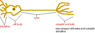
3 Parts to the
Structure
1. Cell body -contain a
large nucleus surrounded by granular cytoplasm
2. Dendrites
-thick branched divisions of the cell body
-bring nerve impulses toward the cell body
3. Axon
-usually a single, longer process that conducts nerve impulses from
the cell body
-terminated at another neuron, muscle, or gland
-may be up to a meter long
Nerve Fiber-common name
for an axon and its myelin sheath
Myelin sheath
-formed by a type of neuroglia cell
-phospholipid segment that wraps around an axon
-protects the axon
-increases the speed of a nerve impulse along the axon
Schwann Cells
-actual cells that from the myelin sheath
-found only in the PNS
Function:
assist in repair of injured axons by providing a tube for the axon or
dendrite to grow
** production of the myelin sheath starts during the 1st year of
life
** amount increases from birth to maturity
-this is the reason that adults react quicker to certain stimuli
Nodes of Ranvier
-segments on the axon that are not myelinated
-gaps in the sheath
![]()
Membrane potentials
-ion concentrations outside of the neuron are very different than
inside the plasma membrane
-neurons have an unequal distribution of potassium and sodium
ions
-K+ concentration is 28 times greater on the inside of the plasma
membrane
-Na+ concentration is 14 times greater on the outside of the
neuron
-inside the membrane are large, nondiffusible, negatively charged
ions
Sodium-Potassium
Pump
-fights osmosis, transports Na+ out and K+ions in in a resting
neuron
-3 Na+ go out for every 2 K+ that are pumped in
-active process that uses ATP energy
Resting Membrane Potential
-neuron with a NET positive charge outside and a NET negative charge
inside the membrane
EXCITABILITY vs. STIMULUS
Excitability
-ability of neurons to respond to stimuli and convert them into
impulses
Stimulus
-any condition in the environment that can alter the resting
membrane
potential
1. If a stimulus is applied, the membranes permeability to Na+
increases at the point of stimulation
2. Na+ rush into the membrane through certain channels. (Na+ are
attracted to the large negative ions in the membrane)
3. More Na+ coming in than being pumped out. Inside shifts from a
negative to positive. (Depolarization)
4. Voltage-gated channels help restore the proper Na+ and K+
concentrations (Repolarization)
(Steps 1-4 occur in a wave motion traveling down the neuron
membrane)
Refractory Period
-time in which the neuron cannot generate another impulse
**under normal conditions each fiber may conduct 10 to 500 impulses
per second
**larger neurons conduct more, up to 2500 per second
Threshold stimulus
-any stimulus strong enough to initiate a nerve impulse
All or None
-once a neuron is stimulated the impulse travels the entire length of
the neuron
-impulse along a myelinated fiber
-myelin sheath inhibits movement of ions
-Nodes of Ranvier allow for action potentials to be generated and
conducted
-ionic current flows through the extra cellular fluid and triggers an
impulse at the next node
-mechanism is the same as continuous conduction BUT the impulse skips
from one node to the next
Valuable to Homeostasis
-speed on impulse greatly increased
- low energy expenditure by Na-K pump because there is not as much
exposed membrane
Synapse
-junction between 2 neurons
-also called synaptic clefts
-essential in homeostasis because of the ability to transmit some
impulses while inhibiting others
-brain diseases and many psychiatric disorders result from bad
synaptic communication
-site that certain drugs effect
2 types of synapses
electrical and chemical
**most synapses in the CNS are chemical
Function:
-neuron secretes neurotransmitters across the synaptic cleft
-post synaptic neuron has receptors to match the transmitter
-when there is a match the impulse will continue
**synaptic vessicles store the neurotransmitters
**drugs trick the receptors, you feel or don’t feel things that
aren’t really happening
ACETYLCHOLINE (ACh)
-most common neurotransmitter
-released by many presynaptic axons in the PNS
-neurotransmitter used to stimulate muscles
1. ACh released from the end bulb--calcium ions trigger the release
of the synaptic vessicles
2. ACh crosses the cleft
3. Fits into the post synaptic receptors
4. Stimulates the neuron--increases the permeability toward Na+
5. Depolarization begins
6. Nerve impulse continues or the muscle contracts
-after 6 months of age, neurons lose their ability to
reproduce
-myelinated peripheral neurons MAY regenerate damaged axons if the
cell body remains intact
-central nervous system neurons are myelinated by
oligodendrocytes-- don’t
help in regeneration
therefore a damaged neuron in the CNS is functionally dead
-1300g
-one of the largest organs
-4 main parts
Brain Stem
-looks like a mushroom stalk
-consist of the medulla oblongata, pons, and mesencephalon
Diencephalon
-consist of the thalamus and hypothalamus
Cerebrum
-looks like the cap of a mushroom
-spread over the diencephalon
-7/8 of the total mass of the brain
-fills most of the cranium
Cerebellum
-inferior to the cerebrum and posterior to the brain stem

Cranial Meninges
-one layer adheres to the cranial bones
-one layer adheres to the brain directly
pia mater--transparent,
fibrous with many blood vessels
Cerebrospinal Fluid
(CSF)
-flows between the 2 meninges, around the brain and spinal cord
-80 ml to 150 ml in the CNS
-clear and watery, contains proteins, glucose and salts
homeostatic functions
1. protection
-shock absorber
-allows the brain to “float”
2. circulation
-delivers nutrients and removes waste
**CSF circulates between the meninges and through
4 ventricles (cavities)
2 lateral ventricles (one in each hemisphere)
1 between and inferior to the lateral ventricles
1 between the inferior brain stem and the cerebellum

-2% of your body weight (brain) uses 20% of the available
O2
-constant supply of glucose energy is needed
-1 to 2 minute interruptions of O2
may lead to death of neurons

Medulla
Oblongata
-continuation of the spinal cord just superior to the foramen
magnum
-contains all tracts of ascending and descending neurons that
communicate information between the brain and spinal cord
-decussation occurs at the
inferior portion
-crossing over of neural tracts
-allows left side of the cerebral cortex to control motor
movements
on the right side of your body
Reticular formation
is a region that passes through the pons,
mesencephelon, diencephalon and into the spinal cord.
-controls consciousness and arousal from sleep
3 reflex centers (vital)
Pons
“bridge”-superior to the medulla oblongata, anterior to the
cerebellum
function:
connect the medulla oblongata to the brain and other parts of the
brain to each other
Mesencephalon
(mid-brain) between the pons and diencephalon
function:
-reflex centers for movements of the eyeball and head in response to
visual stimulus
-reflex centers for movements of the head and trunk in response to
auditory stimuli
-oculomoter nerves
-fine touch nerves
Diencephalon
(2 parts)
-water concentrations
-hormone concentrations
-blood temperature
homeostatic functions
1. regulates autonomic nervous system
2. reception and integration of sensory impulses from viscera
3. coordinates nervous and endocrine system
4. mind over body (stress--heart rate increases)
5. rage and aggression
6. regulates body temperature
7. regulates food intake (hunger and full feelings)
8. thirst
9. sleep patterns
-sits on brain stem and forms the bulk of the brain
Cerebral
cortex
-is the surface of the cerebrum
-folds in the cortex are called
gyri or
convolutions
-shallow grooves between the convolutions are called
sulci
-deep grooves in the cortex are called
fissures
Corpus
callosum--internal bundle of fibers that connect the 2
hemispheres
result from displacement and distortion of neurons at the moment
of impact
1. CONCUSSION
-abrupt but temporary loss of consciousness following a blow to the
head or the sudden stopping of a moving head
-no visible bruising but post traumatic amnesia may occur
2. CONTUSION
-visible bruising of the brain due to trauma and blood leaking from
microscopic vessels
-pia mater is torn
-results in unconsciousness for several minutes to many hours
3. LACERATION
-tearing of the brain , usually from skull fractures of gunshot
wound
-large blood vessels bleed into the brain and can cause cerebral
hematoma, and increased cranial pressure
Lobes and sections of the brain are named after the skull bones
the areas are under
Sensory
Areas
interpret senses
Clinical application: Positron
Emission Tomography
Motor
Areas
Split Brain Concept
anatomically different:
-frontal lobe of the left hemisphere is smaller
-in left handed people the right parietal and occipital lobes are
narrower
functionally different:
-right hemisphere:
left handed, music and artistic awareness, space and pattern
perception, imagination
-left hemisphere:
right handed, language, numerical and scientific skills, sign
language and reasoning

-2nd largest part
-separated from the cerebrum by the transverse fissure
function:
-coordinates subconscious movements of skeletal muscles
-coordinates information from receptors in muscles and tendons with
impulses your motor areas are trying to have you do (ensures smooth
movements of your body)
-maintains posture and equilibrium
-predicts future position of body part during movement
Damaged cerebellum:
-lack of muscle control
-change of speech pattern
-severe dizziness
-disturbances of gait (walking)
|
|
|
|
|
|
|
|
|
|
|
|
|
|
|
|
|
|
|
|
|
|
|
|
|