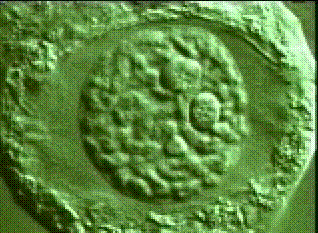
There are three basic planes in which round organs can be sectioned that are useful in reconstructing their three dimensional anatomy. One of these, the transverse or cross section, is made at right angles to the longitudinal axis of the organ. The other two are longitudinal sections and are made parallel to the longitudinal axis of the organ. Radial longitudinal sections are prepared parallel to a radii of the organ, whereas tangential longitudinal sections are made at an angle to a radii of the organ. An exact radial longitudinal section is referred to as a median longitudinal section and is quite instructive in studies of meristems, but extremely difficult to make via free hand sectioning.
For flat plant material, such as leaves and floral organs, the best results can be obtained by sandwiching the plant part between two pieces of styrofoam (or pith or potato) which can be securely grasped. Section the sandwich as you would a more massive piece of plant, then pick out the sections of interest from the water bath after floating the pieces of the sandwich.
There are also three basic planes in which flat plant material can be sectioned. The transverse or cross section is made orthogonal to the longitudinal axis. The sagittal or longitudinal section is made parallel to the longitudinal section and at right angles to the epidermal layers. The paradermal section is made parallel to the epidermis of the organ and is rather difficult to achieve via free hand methods.
Histological stains can either be general, in which case, the generally translucent features of plant cells are dyed some color that makes these features easier to resolve; or specific, in which case, a specific dye that reacts with a particular class of molecules (such as chromatin) is used to help resolve the location of these molecules in the plant material. Often stains can be used in combinations that help localize several classes of molecules in one section.
Since various
plant material will react at various rates with different dyes you must
experiment with each type of tissue to achieve the desired
results.
The experimental variable in most cases is the time you allow the
material
to react with the different dyes. Use several sections for each
histological
procedure. Remove one or two sections from the staining dish
after
regular periods of time have elapsed. Note
in linked dye pages which times give you the best results for specific
material. This will speed up your future observations on similar
material in future exercises.
|
|
|
|
|
|
| Fast Green | Cytoplasm |
| Toluidine Blue | Nuclear Material |
| Basic Fuchsin | Nuclear Material |
| Erythosin B | General Purpose |
Mount your material in a 50% Aqueous solution of Glycerine. You can seal the edges of the coverslip with clear fingernail polish and store your slides in a refrigerator for several weeks without degradation of the stain quality for most dyes.
Elodea, favorite material for observation of many general features of cellular structure, is well suited for review and for emphasizing certain observational techniques and practices. Mount younger leaves of the terminal cluster in water, adaxial side up. Stain similarly age leaves with the available general histological stains. Record your observations on unstained and stained leaves in your exercise sheet. Learn how to calibrate and make linear measurements in Image J, so that you can compare two methods of measuring dimensions of of Elodea in your exercise sheet.
II. Watch this snippet of plant (Algae) cell division or this one, or both! Then complete exercise II in your exercise sheet.
III Plastids
A.
Chloroplasts.
Prepare thin razor blade sections of stems of Pelargonium
(2.03). Direct attention to cells of outer cortex which
should show starch grains in the chloroplasts. Pigment may
be
restricted to a localized region at the periphery. Slide should be
searched to reveal the localized pigment in an equatorial view.
Add a few drops of IKI
at edge of cover glass and observe effect.
B.
Chromoplasts.
Observe pigment-bearing structures in materials
provided -- red pepper, tomato, flower petals, etc. Also make
thin razor blade sections of deep phloem (after peeling) from
carrot root.
C.
Leucoplasts.
Observable in cells of potato tuber immediately
under brown periderm, and in lower epidermis of Zebrina leaf.
Leucoplasts in which starch is deposited may be observed in
potato, and endosperm of cereal grains (split and scrape a
small amount of tissue on a slide). Apply IKI.
Provide an example of each of these plastids in your exercise sheet.
IV. Crystals.
Examine free-hand preparations for crystals of different kinds using bright field and polarized light microscopes.
A. Prismatic -
Ficus leaf (2.05).
B. Raphides -
Yucca
leaf (2.08).
C. Druses - Ginkgo
stem (2.07).
D. Styloids -
mesophyll of Iris leaf (2.08).
E. Cystolith
- epidermis of Ficus leaf (2.05).
Provide at least one example of one of these crystals in your exercise sheet.
V. Compare the structures
visible
with light microscopy with
those
visible in this transmission electron
micrograph
of a plant cell.
Then complete
exercise V in your exercise sheet.