|
How does Confocal Microscope Work?
|
There are some lenses inside the microscope, these lenses focus light from the focal point of one lens to another point. The light from another point in the sample, which is not at the focal point of the lens, but is imaged by the lenses of the microscope. The image of one focal point is not at the same location as the image of the another focal point. Points don't need to be at the focal point of the lens in order for the lens to form an image. So, we want to just look at the one point, that is, the point directly at the focus of the lens. If we put a screen with a pinhole at the other side of the lens system, at the image of the one focal point, then all of the light will pass through this pinhole. However, most of the light from the second focal point is still out of focus at this screen, and gets blocked by the pinhole.
|
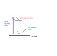
Normally, the sample is completely illuminated by the excited light, so all of the sample is fluorescing at the same time. Of course, the highest intensity of the excited light is at the focal point of the lens, but other parts of the sample do get some of this light and they do fluoresce. This contributes to a background haze in the resulting image. Adding a pinhole combination solves this problem. Because the focal point of the objective lens of the microscope forms an image where the pinhole is, these two points are known as conjugate points. The pinhole is conjugate to the focal point of the lens, making it a confocal pinhole.
|

CLICK IMAGE FOR MORE DETAIL
The confocal microscope simply has two pinholes through which the light must pass before it enters the eye. A laser is used to provide the excited light, or light with very high intensities. Light from the laser must pass through the first pinhole A before it reaches the condenser lens, which focuses it onto the specimen. Light from the specimen passes through the objective lens and is focused through the second pinhole B. Any light coming from the specimen that is not in focus will not pass through the second pinhole B and will not be detected.
|

CLICK IMAGE FOR MORE DETAIL
|
The advantage of the confocal microscope over the normal light microscope is the subtraction of out of focus light so a much clearer image is produced. Any part of a specimen that is blurry cannot be seen with the confocal so the image has no depth.
|
There never is a complete image of the sample, at any given instant, only one point of the sample is observed. The detector is attached to a computer which builds up the image, one pixel at a time. In practice, this can be done perhaps 3 times a second, for a 512x512 pixel image. The limitation is in the scanning mirrors. This uses a high-frequency sound wave in a special crystal to create a diffraction grating, which deflects the laser light. By varying the frequency of the sound wave, the computer changes the angle of the diffracted light, helping scan the sample quickly, allowing 512x480 pixel images 30 times per second. If you want to look at a smaller field of view, confocal microscopes can go even faster, up to 480 frames per second, although 240 frames per second is a good practical limit.
CLICK HERE FOR MORE DETAIL
|
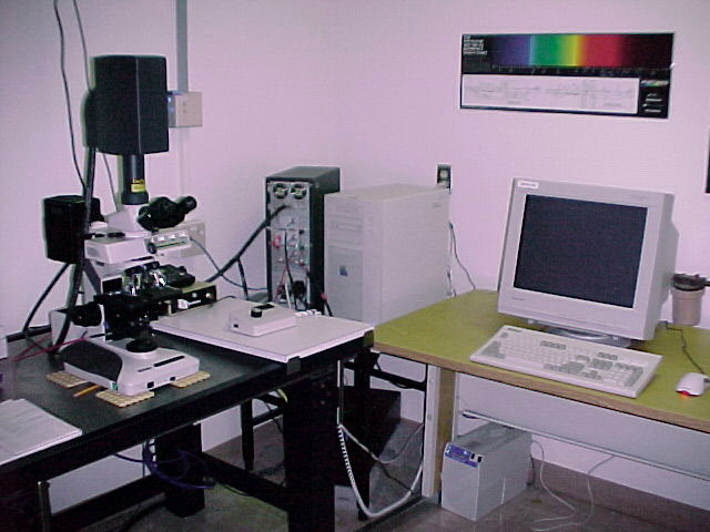
This digital computer (digitizer) transfers the image from the microscope to a computer screen for viewing.
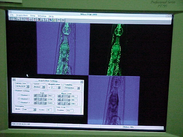
This is an image of a nematode taken at Miami University in Oxford, Ohio
|