Objectives
Study the anatomical features that are involved in the absorption of water, minerals and carbon dioxide.
Plants are autotrophic
organisms that live by converting the electromagnetic energy of light into
chemical energy. Although they are independent from external sources
of chemical energy they do require external sources of water and carbon
dioxide to perform the light and dark reactions of photosynthesis.
In addition plants require external water and essential mineral elements
in order to grow and maintain vital processes.
I. Absorptive processes providing water and mineral elements
A. Carefully remove barley from the soil matrix in which it is growing. Wash the roots with running water. Notice that the extreme tip and more proximal parts of the roots wash clean of soil particles but that in the portion of the root in between these regions the soil particles adhere to the root. Examine these regions under the dissecting microscope. Why do the soil particles adhere to the root? This region corresponds to the region of active absorption of water and mineral elements. Record your observations in the lab exercise sheet.
B. Study the Zea and Pisum roots that have been immersed in a solution of neutral red or a weak solution of eosin for 4, 12, and 24 hours by preparing free hand transverse sections at various places along the longitudinal axis of the root. Where do you find the dye? Is dye penetration into the interior of the root uniform throughout the longitudinal axis? What do your observations tell you about the absorption function of roots? Locate the endodermis by staining the casparian strip in one of the root samples. Record your observations in the lab exercise sheet.
C. Some epiphytic plants lack roots (Tilandsia) or have aerial roots (many Bromiliads and Orchids) which have a multiple epidermis called a velamen. These plants absorb water and elements from rainfall and dews which fall or condense on their aerial surfaces. Record your observations on one of the following examples in the lab exercise sheet.
1. Make observations using the dissecting microscope and free hand sections of Tilandsia leaves and Orchid roots that are dry and that have been exposed to neutral red or a weak solution of eosin. Comparison of the anatomy of these structures subject to the two conditions will reveal how water and elements are absorbed in these organs.
2. Perform similar
experiments with Pinus fascicles. Pay special
attention to what happens in the region of
scale leaves surrounding the proximal part of the
fascicles.
II. Absorptive processes involving gases
The CO2 utilized in the dark reaction of photosynthesis must diffuse into the chloroplasts from the external atmosphere. The greater the exposure of surface area of chlorenchyma cells, the more rapid this will occur. On the other hand, however, the greater the exposed surface area of these cells, the more rapid the loss of water will be to the atmosphere. Plants have evolved specialized guard cells within the aerial epidermis to regulate this gaseous exchange, necessary to their existence. Record your observations in the lab exercise sheet.
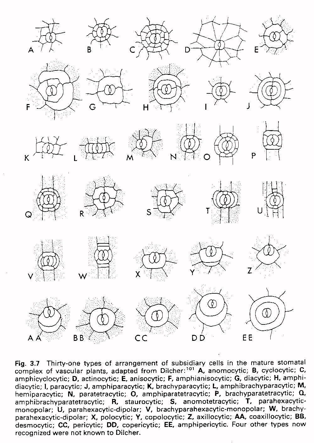
A. Make epidermal impressions of two species for which fresh leaf material is provided using the cellulose acetate peel technique. Determine what type of stomata these species have. The features that these different types share in common reveal the necessary functional aspects of stomata, whereas the differences between them will give you an idea of the biological variation that occurs within these functional constraints.
In plant organs, like woody stems, that lack chlorenchyma tissue, oxygen, necessary for respiration, may become limiting. Specialized openings in the phellem, termed lenticels, facilitate gaseous exchange through this otherwise impervious tissue. This gaseous pathway can be illustrated by blowing air into the base of a twig that has been immersed in a beaker of water.
B. Study the geometry
and distribution of lenticels on the
twigs you have collected. Prepare free hand
sections through these lenticels to determine their relationship
with living internal tissues, the surrounding impervisous cork tissue,
and the phellogen, or cork cambium. Can
you recognize filling or complementary cells and closing cells within your
lenticel preparation? Record
your observations in the lab exercise sheet.
Material
Prepared Slides Fresh
Coffea (5.17)
2 wk old plants of
Dianthus (7.02)
Nerium (7.03)
Zea
Zea (3.07)
Pisum
Pinus (3.09,3.095)
Sambucus (5.05,5.051,5.052)
Tilandsia
Tilia (3.05,3.055)
Orchid roots
Betula (7.07,7.071)
Pinus fascicles
Leaves of:
Ranunculus
Kalanchoe
Dianthus
Bignonia
Nerium
Zea
Twigs of:
Zelkova
Prunus
Quercus
Pinus
Other:
Cellulose acetate
Acetone