I. Initial material deposited during cell plate formation
A. Initially rich in pectins (middle lamella), amount of cellulose increases over time
B. Subsequent material deposited through plasmalemma to space between middle lamella and plasmalemma
II. Primary function is support
III. Many other diverse secondary functions dependent of pattern and type of materials
A. Determines cell shape
B. Cell to cell communication
C. Protection from mechanical damage and disease organisms
D. Recognition and binding of external molecules
E. Control of water pathways (lignin)
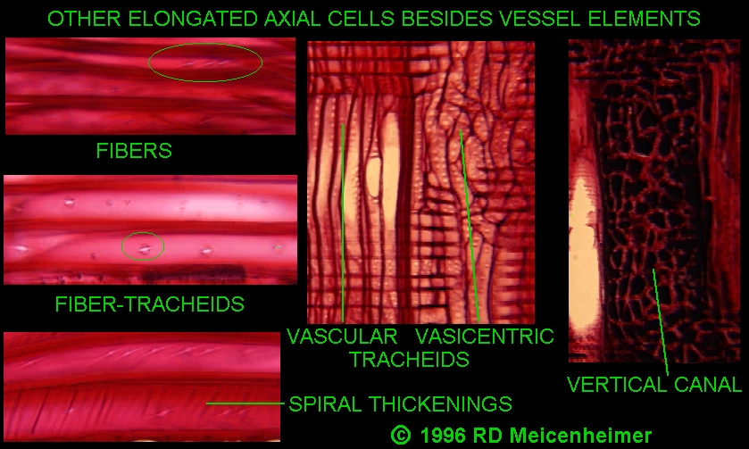
IV. Extracellular matrix of all eukaryotes share two common structural elements
A. Fibers
1. Long, semicrystalline elements
2. Provide resistance to
stretching and other tensile forces
B. Network
1. More or less elastic, interlocked
assembly of branched molecules
2. Hold fibers in place
3. Provide resistance to
compressive forces via trapping and retarding flow of water molecules
V. Physical properties of extracellular matrices determined by
A. Kinds of fiber and network molecules present
B. Degree of crosslinking of fibers via network molecules
C. Amount of trapped water
D. Types of additional material
within the matrix
1. Lignin in plant cell walls
DETAILS OF PLANT EXTRACELLULAR MATRIX COMPONENTS
I. Fiber
A. Cellulose = long straight chains of B1,4-linked glucose
1. 20 - 50% dry weight
2. 15% volume
B. Form Micofibrils with quasi-crystalline
structure (10-85 nm diameter)
via hydrogen bonding between
cellulose molecules
1. Primary
Cell Wall
2. Secondary
Cell Wall
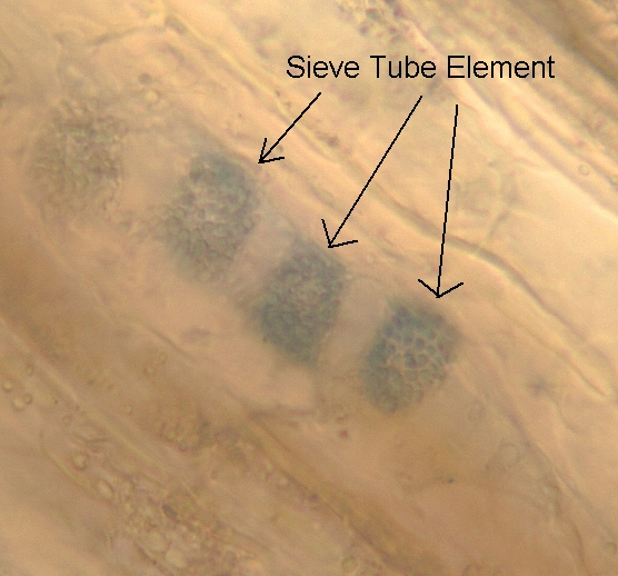
C. Callose = long chains of B1,3-linked glucose
1. Sieve tube elements and wounded plant cells
II. Network
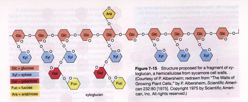
A. Hemicellulose = branched chains of glucose + other sugars
1. 20% dry weight
2. Form sheathing layers
around microfibrils via hydrogen bonds
3. May be neutral or acidic
inl pH range of cell wall
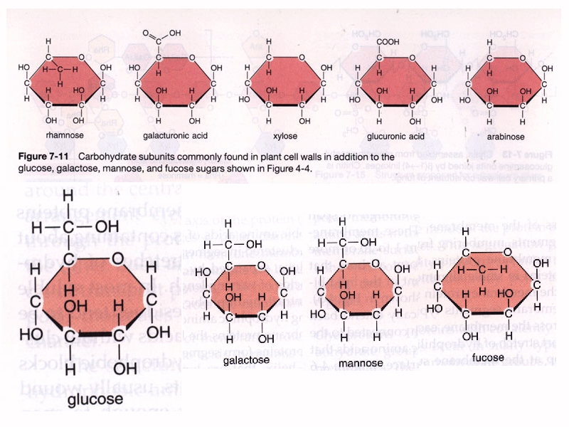
B. Pectins = branched chains of polysaccharides with galacturonic acid or galacturonic acid + rhamnose backbones
1. 25 - 30% dry weight
2. Form stable gels of various
viscousity via water trapping
3. Highly acidic in pH range
of cell walls = electrostatic bonds with other molecules
(Interactions with Ca+2 might play a linkage role?)
III. Other network components
A. Various enzymes
1. Transferases involved in
moving sugar groups from precusers to network components
2. Hydrolytic enzymes that
remove sugar groups and create binding sites in network components
3. Peroxidases, phosphatases,
proteases, oxidases, and reductases
a. Some of these hydrolyze
molecules of cell walls of invading fungi and bacteris
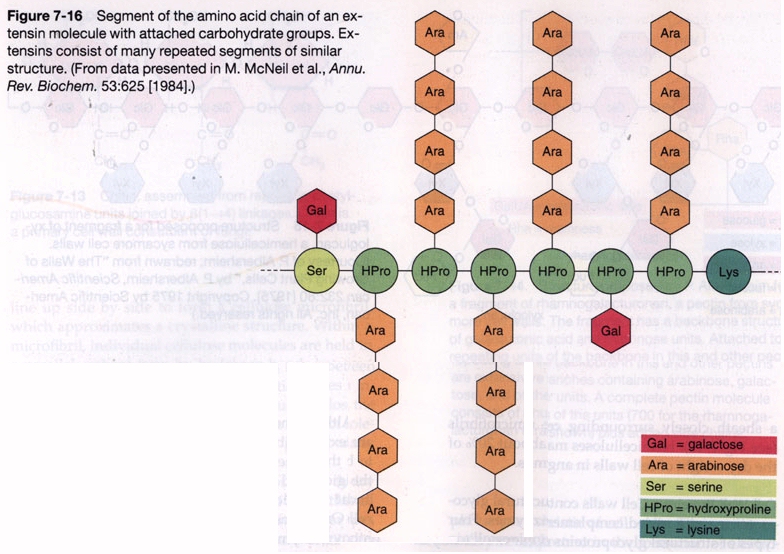
B. Structural Glycoproteins rich in hydroxyproline or glycine
1. Extensins confer rigidity to cell wall via ionic attractions with negatively charged network components and covalent bonds with other extensins and pectins
C. Lectins
1. Soluble glycoproteins with
multiple binding sites to cross link carbohydrate groups of other network
components
2. Can bind with chitins
of invading fungi
3. Part of recognition mechanism
between root hairs and Rhizobium bacteria in legume roots
D. Silica important component of cell walls of grasses and Equisitum
E. Lignin
1. Dense, insoluble substance
formed by complex alcohols covalently linked into branched network
2. Lignification makes cell
walls hydrophobic which is important in function of endodermis of monocot
roots and vascular cells
F. Suberin, Cutin, and Waxes
1. Insoluble substances formed
by complex fatty acids covalently linked polymers
2. These substances make
cell walls hydrophobic which is important in function of endodermis
of dicot roots, epidermal cells, and periderm (bark)
cells
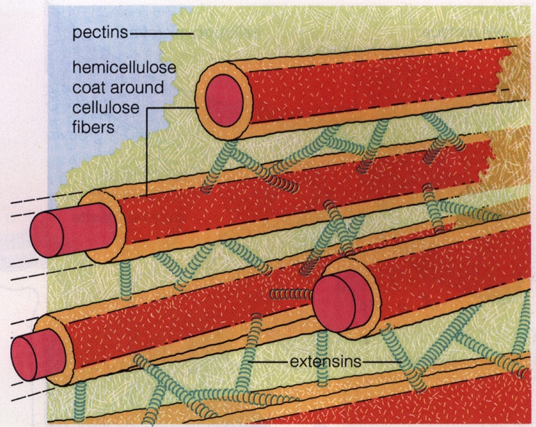
IV. Structure of cell wall only partially understood through enzymatic digestion studies
A. Cellulose microfibrils surrounded by hydrogen-bonded hemicellulose sheath
B. These units cross-linked via hydrogen-bonded pectins
C. Cross-linkage may be reinforced via extensins and other glycoproteins
D. This network is open enough to admit water, ions, and small molecules but has great resistance to stretching and compression
E. Proportions of cellulose, hemicellulose, and pectins varies between
1. Layers within the
cell wall
2. Different types
of cells
3. Different species
of plants
F. Size and orientation of cellulose microfibrils varies between
1. First formed primary cell wall
a. Some cells have thin primary cell walls
b. Some cells have thick primary cell walls
2. Latter formed secondary cell wall
a.
Composed of three layers (S1,S2,S3)
b. Most cells with secondary cell walls undergo apotosis and have
no cytoplasm in lumen at maturity
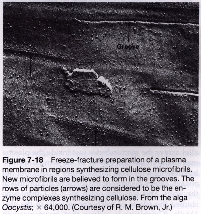
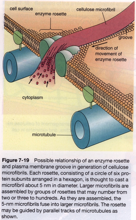
V. Synthesis of cellulose microfibrils occurs at the plasmalemma
B. Microtubules thought to somehow be involved in the orientation of microfibrils
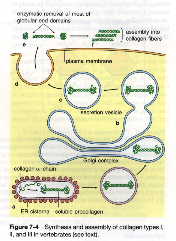
VI. Synthesis of network molecules
A. Initial precursors via Golgi apparatus -> vesicles to plasmalemma
B. Final synthesis and linkages
via enzymes in extracellular matrix
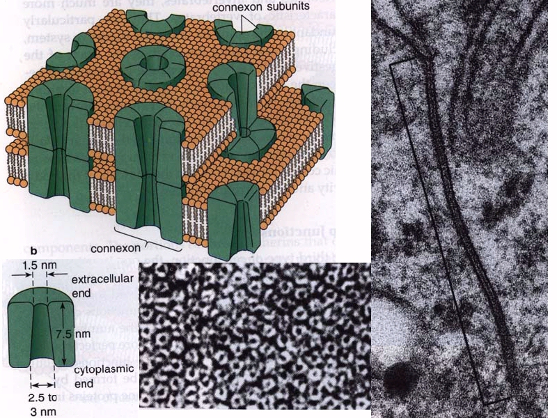
COMMUNICATION BETWEEN PLANT CELLS
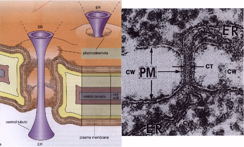
I. Via Plasmadesmata in primary cell wall
A. Plasmalemma continuous between adjacent cells
B. Endoplasmic reticulum between adjacent cells connected via central tubule
C. Upper limit of free movement = 700 daltons
D. Antibodies against connexin proteins (Animal gap juctions) react with plasmadesmata
E. Evidence that movement regulated by divalent cations
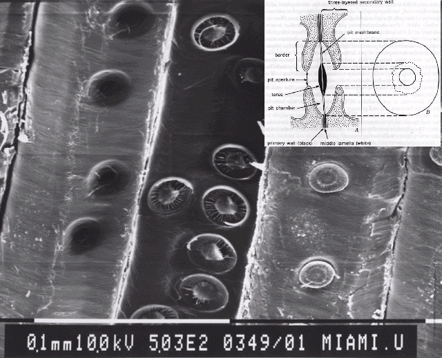
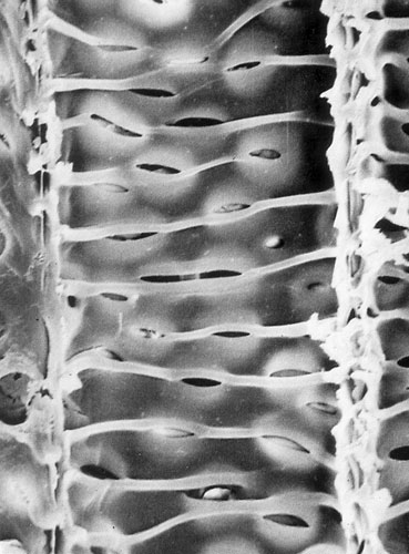
II. Via Pits in secondary cell wall
A. Regions where secondary cell wall is not deposited
B. Two continuous regions which can vary independently in shape and size
1. Pit canal = adjacent to cell lumen or plasmalemma
2. Pit chamber = adjacent to primary cell wall
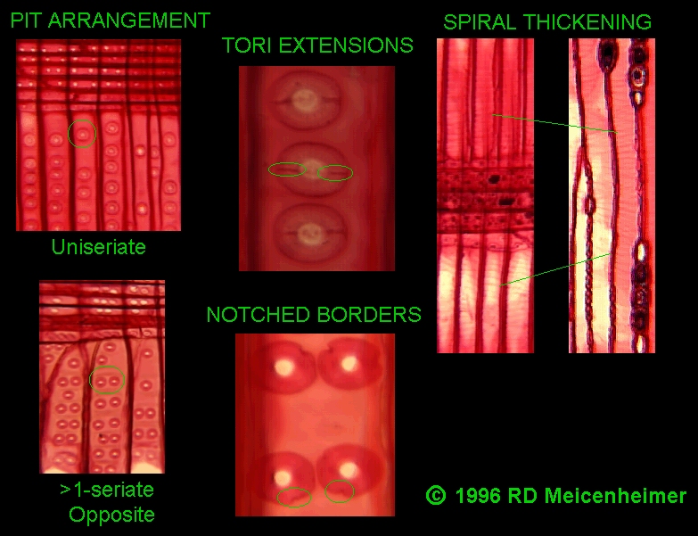
3. Different cell types have different types of pits
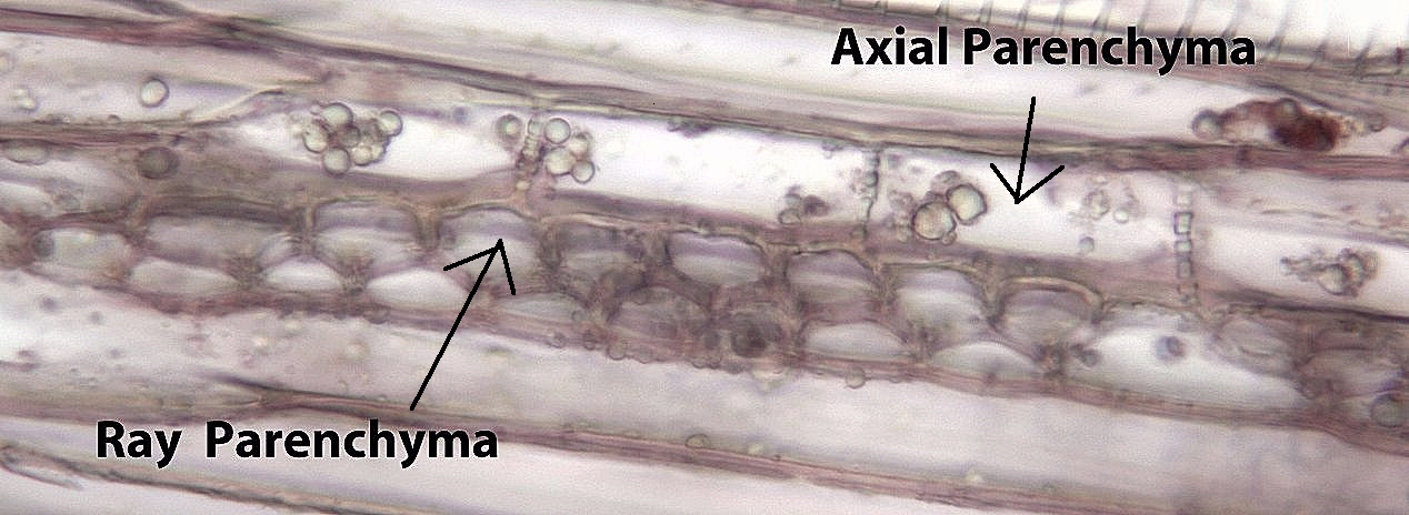
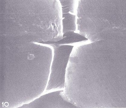
a. Simple pits
1. Pit chamber = Pit canal in size (SEM View) (Ray & Axial Parenchyma)
2. Single pit chamber connected with branching pit canal (ramiform) (Pyrus Brachysclereid)
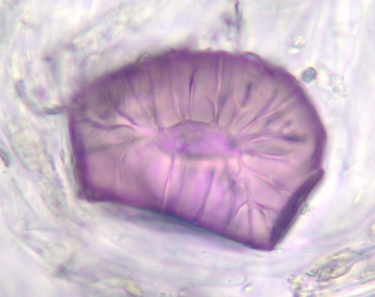
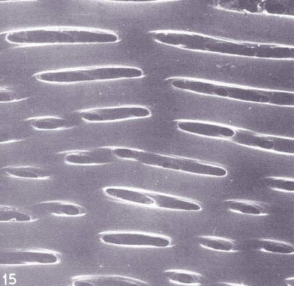
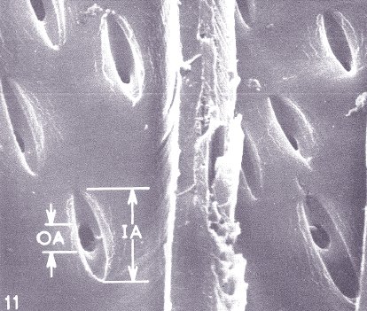
b. Bordered pits: Pit canal <> Pit chamber in size and shape
1. Circular pit chamber and pit canal (SEM View) (Tracheids)
2. Elliptical pit chamber and pit canal (SEM View A) (SEM View B) (Vessels)
3. Circular pit chamber with slightly larger elliptical pit canal (Tracheids)
4. Circular pit chamber with much larger elliptical pit canal (SEM View) (Fiber-Tracheids)
5. Very small circular pit chamber with large eliptical pit canal (SEM View) (Fibers)
C. Pit pairs are
formed via alignment of pits of two adjacent cells